Gaining all weight back! Failed band results!
on 2/25/12 12:22 am - ND
ETA: Well, crap..it IS Saturday ...doubt your doc is open....but if your doc is like mine, he would want you to call him in a situation like this.
5.0 cc in a 10cc lapband (four fills) 1 unfill of .5cc on 5/24/2011.
.5 fill March 2012. unfill of .25cc May 2012. Unfill of .5cc June 2014.
Still with my lapband with no plans for revision. Band working well since
last small unfill.
HW: 267lbs- size 22-24 LW:194lbs CW:198lbs Size 14-16
on 2/29/12 5:24 am, edited 2/29/12 5:25 am - Califreakinfornia , CA
The esophagus functions solely to deliver food from the mouth to the stomach where the process of digestion can begin. Efficient transport by the esophagus requires a coordinated, sequential motility pattern that propels food from above and clears acid and bile reflux from below. Disruption of this highly integrated muscular motion limits delivery of food and fluid, as well as causes a bothersome sense of dysphagia and chest pain. Disorders of esophageal motility are referred to as primary or secondary esophageal motility disorders and categorized according to their abnormal manometric patterns. See the images below.
Anatomy
The tubular esophagus is a muscular organ, approximately 25 cm in length, and has specialized sphincters at proximal and distal ends. The upper esophageal sphincter (UES) is comprised of several striated muscles, creating a tonically closed valve and preventing air from entering into the gastrointestinal tract. The lower esophageal sphincter (LES) is composed entirely of smooth muscle and maintains a steady baseline tone to prevent gastric reflux into the esophagus.
The body of the esophagus is similarly composed of 2 muscle types. The proximal esophagus is predominantly striated muscle, while the distal esophagus and the remainder of the GI tract contain smooth muscle. The mid esophagus contains a graded transition of striated and smooth muscle types. The muscle is oriented in 2 perpendicular opposing layers: an inner circular layer and an outer longitudinal layer, known collectively as the muscularis propria. The longitudinal muscle is responsible for shortening the esophagus, while the circular muscle forms lumen-occluding ring contractions.
Esophageal peristalsis
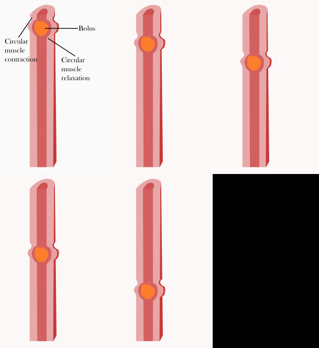
The coordination of these simultaneously contracting muscle layers produces the motility pattern known as peristalsis. Peristalsis is a sequential, coordinated contraction wave that travels the entire length of the esophagus, propelling intraluminal contents distally to the stomach. The LES relaxes during swallows and stays opened until the peristaltic wave travels through the LES, then contracts and redevelops resting basal tone. Low peristaltic amplitudes normally occur at the transition zone between the striated and smooth muscle portions; however, the peristalsis is uninterrupted.
Primary peristalsis is the peristaltic wave triggered by the swallowing center. The peristaltic contraction wave travels at a speed of 2 cm/s and correlates with manometry-recorded contractions. The relationship of contraction and food bolus is more complex because of intrabolus pressures from above (contraction from above) and the resistance from below (outflow resistance).
The secondary peristaltic wave is induced by esophageal distension from the retained bolus, refluxed material, or swallowed air. The primary role is to clear the esophagus of retained food or any gastroesophageal refluxate.
Tertiary contractions are simultaneous, isolated, dysfunctional contractions. These contractions are nonperistaltic, have no known physiologic role, and are observed with increased frequency in elderly people. Radiographic description of this phenomenon has been called presbyesophagus.
Esophageal motility disorders
Esophageal motility disorders are not uncommon in gastroenterology. The spectrum of these disorders ranges from the well-defined primary esophageal motility disorders (PEMDs) to very nonspecific disorders that may play a more indirect role in reflux disease and otherwise be asymptomatic. Esophageal motility disorders may occur as manifestations of systemic diseases, referred to as secondary motility disorders.
Esophageal motility disorders are less common than mechanical and inflammatory diseases affecting the esophagus, such as reflux esophagitis, peptic strictures, and mucosal rings. The clinical presentation of a motility disorder is varied, but, classically, dysphagia and chest pain are reported. In 80% of patients, the cause of a patient's dysphagia can be suggested from the history, including dysmotility of the esophagus. Before entertaining a diagnosis of a motility disorder, first and foremost, the physician must evaluate for a mechanical obstructing lesion.
Esophageal motility disorders discussed in this article include the following:
- Achalasia
- Spastic esophageal motility disorders, including diffuse esophageal spasm (DES), nutcracker esophagus, and hypertensive LES
- Nonspecific esophageal motility disorder (inefficient esophageal motility disorder)
- Secondary esophageal motility disorders related to scleroderma, diabetes mellitus, alcohol consumption, psychiatric disorders, and presbyesophagus
Esophagus
After food is chewed into a bolus, it is swallowed and moved through the esophagus. Smooth muscles contract behind the bolus to prevent it from being squeezed back into the mouth. Then rhythmic, unidirectional waves of contractions will work to rapidly force the food into the stomach. This process works in one direction only and its sole purpose is to move food from the mouth into the stomach.[2]
In the esophagus, two types of peristalsis occur.

- First, there is a primary peristaltic wave which occurs when the bolus enters the esophagus during swallowing. The primary peristaltic wave forces the bolus down the esophagus and into the stomach in a wave lasting about 8–9 seconds. The wave travels down to the stomach even if the bolus of food descends at a greater rate than the wave itself, and will continue even if for some reason the bolus gets stuck further up the esophagus.
- In the event that the bolus gets stuck or moves slower than the primary peristaltic wave (as can happen when it is poorly lubricated), stretch receptors in the esophageal lining are stimulated and a local reflex response causes a secondary peristaltic wave around the bolus, forcing it further down the esophagus, and these secondary waves will continue indefinitely until the bolus enters the stomach.
Esophageal peristalsis is typically assessed by performing an esophageal motility study.
Good Luck with your research and if there is anything I can help you with please feel free to ask me.
Lisa
www.obesityhelp.com/group/failed_lap_bands/

 ... Anyway, it really sounds like your doctor should have used this smaller band on a smaller person. I am a 5'3" female with a small frame and my doc decided to use the smaller 10mm band on me ( what size is yours?). because of my inside anatomy.
 Unless you are a smaller than average male, my doctor would not have used a smaller band on you.
Btw, I was banded about 18 months ago and lost about 40 lbs quickly and the rest slowly since then...if you average it over a 6 month period, I am losing 1-2 lbs a month.
5.0 cc in a 10cc lapband (four fills) 1 unfill of .5cc on 5/24/2011.
.5 fill March 2012. unfill of .25cc May 2012. Unfill of .5cc June 2014.
Still with my lapband with no plans for revision. Band working well since
last small unfill.
HW: 267lbs- size 22-24 LW:194lbs CW:198lbs Size 14-16
on 2/29/12 3:52 am - ND
on 2/29/12 5:41 am - Califreakinfornia , CA
Did he inform you of his unauthorized experimentation or did you sign an informed consent for his little project ?


