Images from my upper GI study

As some of you know, my surgeon's NP thinks my band has slipped. Getting this confirmed (or ruled out) has turned into a ridiculous ordeal (see my OH blog if you're curious). On Tuesday I had an upper GI study done at our local hospital. On Wednesday my PCP's nurse called me with the results: NO MENTION of my band's position, even though "band slip" was written on the doctor's order as the reason for the test. The only reference to my band is information that I verbally gave the x-ray tech and which couldn't possibly be determined by x-ray. Now I'm trying to get an amended report that at least describes the position of my band.
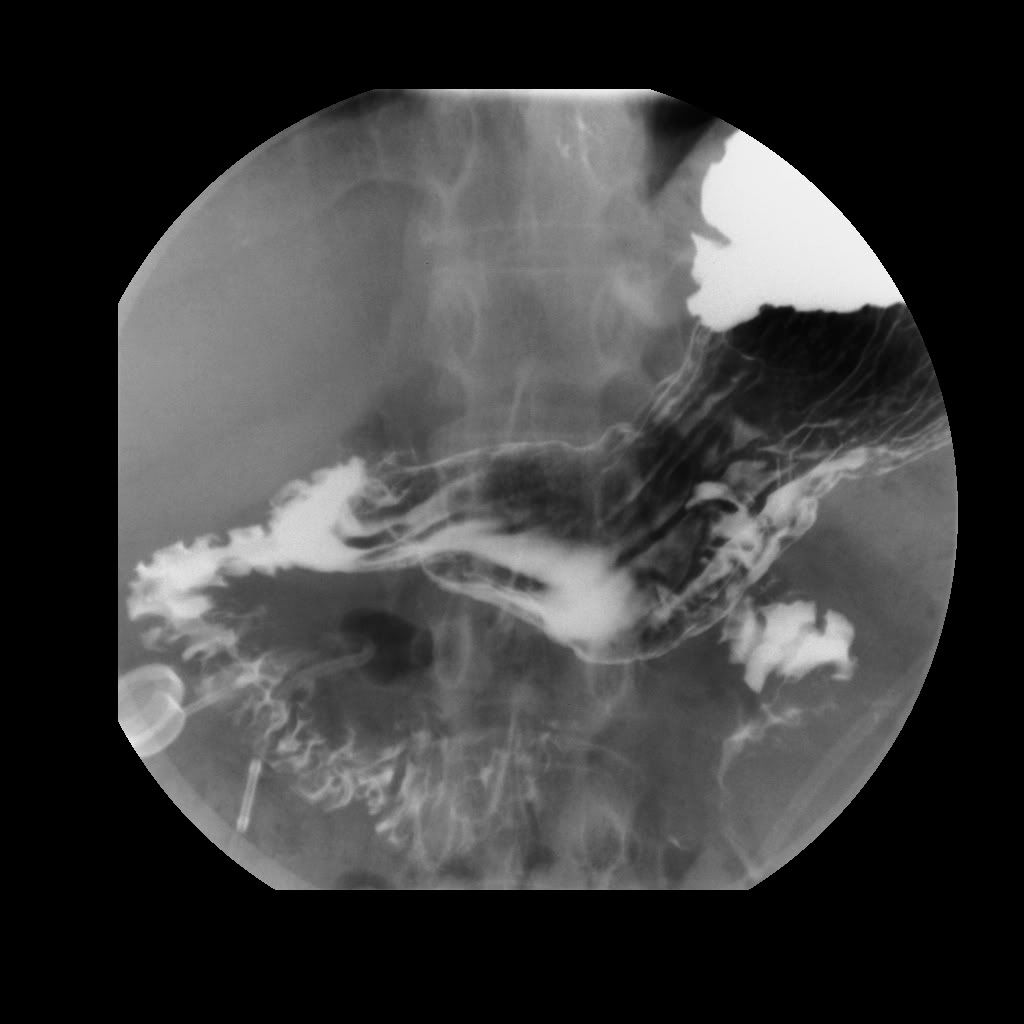
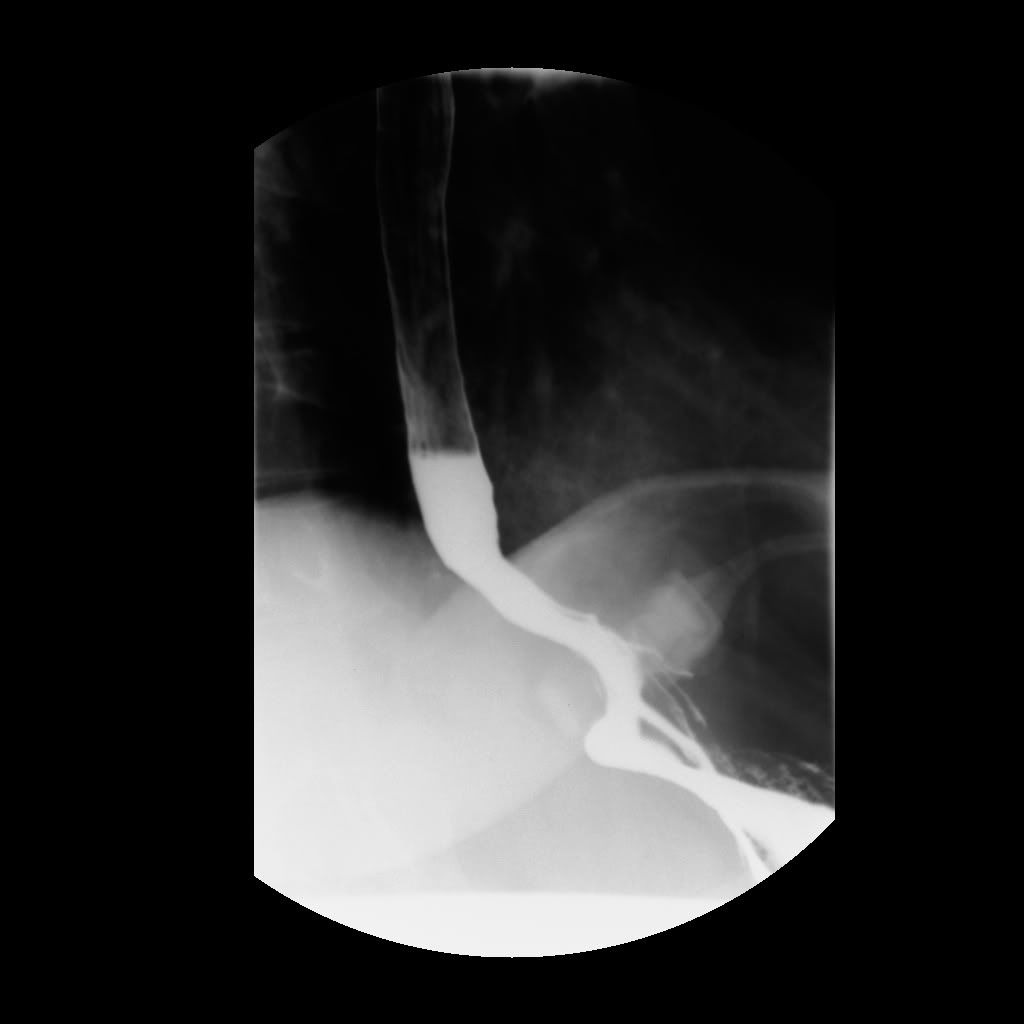
This morning I picked up my own copy of the radiologist's report and a CD of images from my upper GI. Most of the images don't mean much to me, not only because I don't have medical training but because the images were "snapped" at many different angles as I turned around and around on the x-ray machine. In one image it looks like my esophagus is dilated, but that might just be from the pressure of the barium going through it, and in another image it looks normal (that is, not dilated). My port showed up vividly in several shots. I could only find my band in two shots. In one of them, my band seems to be too far up my esophagus.
As bummed out as I am about this whole thing, it's really cool to have these images of my innards. (I just love that kind of thing!) Here's a shot with my port and tubing in the lower lefthand corner.
And here's a shot showing my band on the lower righthand side:
Jean
Jean McMillan c.2009-2013 - Always a bandster at heart
author of Bandwagon (TM), Strategies for Success with the Adjustable Gastric Band & Bandwagon Cookery. Bandwagon for Kindle now available on Amazon. Read my blog at: jean-onthebandwagon.blogspot.com
![]()


(deactivated member)
on 7/23/09 2:36 am - Des Moines, IA
on 7/23/09 2:36 am - Des Moines, IA
Cool pictures Jean. I sure hope you get an answer soon. What has your band Dr. said about these images? Keep us updated!


So sorry you're having trouble Jean. But I'm glad you posted these pictures - very interesting to see.
I seem to recall that Sandy R. posted some pictures of x-rays a while back. My recollection was that there was one scan of a perfectly positioned band - no slip, dilation, etc. - and I have to say, it looked remarkably like your second photo. (not that I know anything at all about it). Check on some of her older posts, and you might find it.

Height - 5'6" French MIDband: 13 fills, current 8.7ccs - capacity 12 ccs


Most Active
Recent Topics
OH - why aren't my posts being approved? Did I do something wrong?
FluffyPuff · 2 replies · 1143 views
FluffyPuff · 2 replies · 1143 views

OH2023 National Conference: Early Bird Tickets On Sale For A Limited Time! SAVE $30!
Member Services · 0 replies · 978 views
Member Services · 0 replies · 978 views

